
When you’re taking a test or enjoying a cup of coffee, certain parts of your brain are extra busy. An fMRI allows us to capture some of this activity.
Functional resonance imaging (fMRI) has revolutionized the study of the mind. This advanced neuroimaging technology allows researchers and physicians to safely, painlessly, and noninvasively observe the brain’s activity.
An fMRI scan can be used for a variety of purposes, from monitoring Parkinson’s disease to seeing how a certain medication works in the brain.
If you’re scheduled for an fMRI, understanding what it is and how it works can help you feel more comfortable.
Invented in the early 1990s, functional magnetic resonance imaging (fMRI) is a type of noninvasive brain imaging technology that detects brain activity by measuring changes in blood flow.
An fMRI can reveal what part of the brain is active during specific functions, such as lifting your arm or even just thinking about something. Researchers and physicians can use this information to better understand, diagnose, monitor, and treat various conditions.
An fMRI is based on the same technology as magnetic resonance imaging (MRI) — a scanning tool that uses powerful magnetism and radio waves to produce images of the body. However, there are important differences between the two.
An MRI takes images of your brain’s structure — it can see cysts, tumors, bruising, bleeding, and structural abnormalities.
When you get an MRI scan, the scan is looking to make sure everything is the right size or is in the right place. For instance, are there any signs of damage, such as bruising? If so, there might be a concussion.
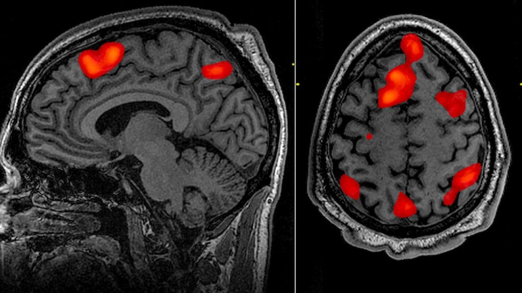
In contrast, an fMRI takes images of your brain’s activity while it’s performing a particular function. It can even “see” your thoughts and feelings. An fMRI is essentially creating a functional map on top of the brain images.
During an fMRI scan, you might be asked to perform a task, like lift your arm or think of the word “summer.” These tasks activate your brain so the fMRI can measure your brain’s activity.
Other times, you might be asked to just relax in the scanner — this is known as a resting state (rs) fMRI. An
An fMRI uses a powerful magnetic field — tens of thousands of times stronger than the Earth’s magnetic field — to detect activity in different parts of the brain.
What does it mean when a brain area is “more active?” And how does the fMRI detect this activity?
A brain area is considered more active when its neurons start sending out more electrical signals than they did before. For instance, if a certain brain area is more “active” when you raise your leg, then that part of the brain is considered responsible for that movement.
An fMRI indirectly measures this electrical activity by detecting changes in oxygen levels in the blood. This is called the blood-oxygen-level-dependent (BOLD) response.
Here’s how this works:
When neurons become more active, they require more oxygen from red blood cells. To achieve this, they widen the surrounding blood vessels to allow for more blood flow. Thus, when the neurons are more active, the oxygen concentration goes up, too.
Oxygenated blood produces fewer field disturbances than deoxygenated blood which allows the neurons’ signal (which is actually hydrogen in water) to last longer. So when the signal stays longer, the fMRI knows that there’s more oxygen in that area, and therefore more activity.
This activity is color-coded in the resulting fMRI images.
fMRIs are used widely in both clinical and research settings. This technology allows us to better understand how the brain works in both healthy and diseased conditions.
In clinical practice, most fMRIs are performed soon after a diagnosis. The resulting brain images can help your healthcare team decide on a treatment plan and whether surgery might be a good option.
An fMRI scan is also commonly given right before brain surgery to help the neurosurgeon prepare.
An fMRI can be used to:
- diagnose conditions
- plan for a surgery or other treatments
- detect abnormalities
- see which brain regions are responsible for important functions
- evaluate the cognitive effects of diseases and injuries, such as epilepsy, concussion, or cancer
- determine drug efficacy
- help with drug development
- understand brain disorders
- examine how memories are formed
- observe how the brain handles critical functions like thinking, emotional responses, learning, movement, sensation, or speech (called brain mapping)
- look for disease biomarkers
- monitor therapy
An fMRI might also be used to detect the following:
- epilepsy
- concussion
- post-concussion syndrome
- mental illnesses, such as schizophrenia
- neurological conditions, such as Alzheimer’s disease and Parkinson’s disease
- tumors
- pain
fMRI is often used to study healthy brains as well. In a small-scale
In the first experiment, the participants were told their chances of winning at the same time they were given the chance to gamble. In a second experiment, the chance was presented before the gambling opportunity.
The findings show that when the participants knew the odds prior to the gambling opportunity (and kept this information in mind), they had more control of their risk-taking behaviors. This was shown by brain activation in regions associated with control and conflict. The participants showed faster reaction times and better performance.
An fMRI unit involves a flat table that slides into a large cylinder-shaped tube surrounded by a circular magnet. “Open” fMRI machines are open on the sides.
A typical fMRI lasts between 45 and 55 minutes.
Before your fMRI, you’ll get instructions on how to prepare and what to expect.
Before the scan
There are no major preparations before an fMRI scan, as there are no injections or invasive procedures.
However, you’ll need to remove your phone, jewelry, glasses, coins, or anything metal (these belongings will likely be put into a safe locker). Medication patches may also need to be removed as the metal in the patch could heat up during the fMRI scan. Keep another one on hand to apply after the procedure.
Any medical implants or devices (such as a stent) may have to be removed prior to the fMRI scan as well.
If you are pregnant or have any health problems, allergies, problems with lying on your back, or claustrophobia (fear of confined spaces), talk with your doctor to help you come up with a plan.
During the scan
During the fMRI scan, you will lie face-up on a flat scanning table that’s rolled into a long, tubular magnet. You may be given instructions, such as squeezing your right hand or thinking of certain words. These activities are color-coded on the brain images, allowing the doctor to see a map of your brain activity.
The process is painless, but some people may feel bothered by the small space or loud noises the machine makes. A typical fMRI lasts between 40 and 55 minutes.
After the scan
Once your fMRI scan is complete, the technologist will slide the scanning table out of the machine and help you up. You can collect your belongings and leave the scanning area. Your doctor will receive a report of your fMRI results which will be used to plan your care.
Before the fMRI was invented, the only way to identify the brain’s motor or language skills center was to stimulate the brain during an invasive procedure, such as surgery. With fMRI, we now have a safe, painless, and noninvasive way to see brain activity.
If you’d like more information, there are plenty of online sites and videos explaining fMRI. This video gives a detailed description of how fMRI works.